
Sparking Life: Bioelectric Communication During Zebrafish Development
Bentley Smallwood and Rachel Lukowicz-Bedford


Transcript
Overview
A colorful digitally drawn comic. The artist uses block and bubble letters, background textures and background stars to emphasize information and add texture. The transcript uses quotes to indicate when text is written in the comic, other text is description. Text has been lightly edited to add additional punctuation.
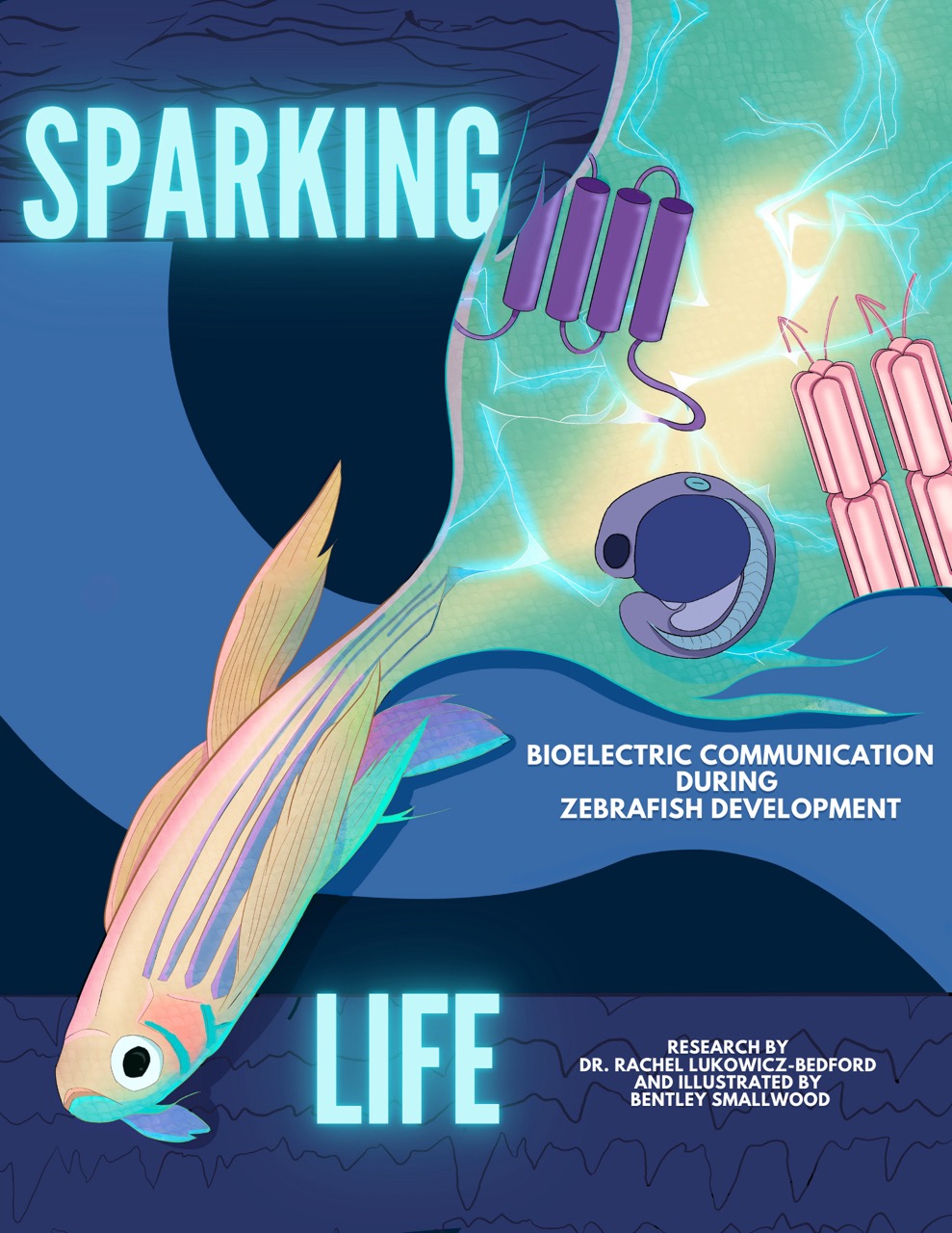
Cover
Sparking Life
Bioelectric communication during zebrafish development
Research by Dr. Rachel Lukowicz-Bedford and illustrated by Bentley Smallwood
Cover description: The title is written in bold, glowing capital letters. The top and bottom of the page are covered with dark blue bands. Small black lines run through the bands, evoking the ripples of water or the lines on a seismograph or EKG. A larger-than-life iridescent zebra fish splashes across the cover. It’s a small fish that is long and thin, with many feathery fins. It has five stripes along the length of its body, wide set eyes, and its mouth points upwards as if it is frowning. Inside its elongated tail are diagrams of cellular structures. Strands of lightning run through the tail.
Page 1
“Thank you to University of Oregon Zebrafish Lab” The text is written in large bubble letters on a background of clear blue water and large leaves. “Zebrafish Lab” is written on two zebrafish.
Page 2
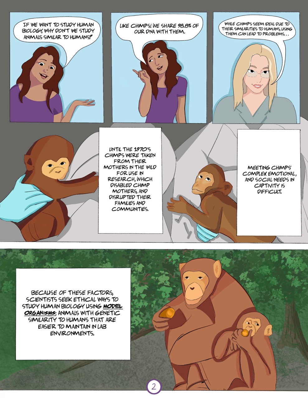
Panel 1: “If we want to study human biology, why don’t we study animals similar to humans?” A young woman with long brown hair and tan skin addresses the reader.
Panel 2: “Like chimps! We share 98.8% of our DNA with them.”
Panel 3: “While chimps seem ideal due to their similarities to human, using them can lead to problems…” A woman with shoulder length blonde hair, pale skin and glasses picks up the narration.
Panel 4: “Until the 1970s chimps were taken from their mothers in the wild for use in research, which disabled chimp mothers, and disrupted their families and communities. Meeting chimps’ complex emotional, and social needs in captivity is difficult.” Two baby chimps hug their human caretakers. The chimps are drawn in full color, but the scientists are drawn in greyscale except for clinically blue latex gloves.
Panel 5: “Because of these factors scientists seek ethical ways to study human biology using model organisms: animals with genetic similarity to humans that are easier to maintain in lab environments.” A young chimp and a mother chimp crouch together in the woods eating fruit.
Page 3
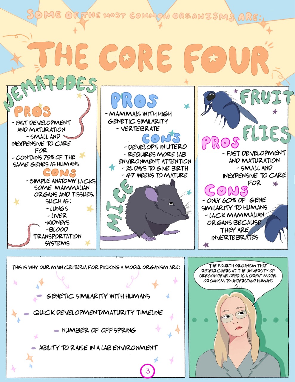
Page title: “Some of the most common organisms are: The Core Four” The title is in large bubble letters; the background is a giant yellow star with smaller stars dotted around the page.
Panel 1: “Nematodes:
Pros:
- Fast development and maturation.
- Small and inexpensive to care for.
- Contains 75% of the same genes as humans.
Cons
- Simple anatomy lacks some mammalian organs and tissues such as:
- Lungs
- Liver
- Kidneys
- Blood transportation systems”
Long thin worm-like organisms decorate the panel.
Panel 2:
“Mice:
Pros
- Mammals with high genetic similarity.
- Vertebrate.
Cons
- Develops in utero.
- Requires more lab environment attention.
- 21 days to give birth.
- 4-7 weeks to mature.”
A small mouse crouches in the corner of the panel.
Panel 3:
“Fruit Flies:
Pros
- Fast development and maturation.
- Small and inexpensive to care for.
Cons
- Only 60% of gene similarity to humans.
- Lack mammalian organs because they are invertebrates.”
Two large fruit flies hover in the panel.
Panel 4: “this is why our main criteria for picking a model organism are:
- Genetic similarity with humans.
- Quick development/maturity timeline.
- Number of offspring.
- Ability to raise in a lab environment.”
Panel 5: The blonde scientist frowns at the reader, “The fourth organism that researchers at the University of Oregon developed as a great model organism to understand humans is…”
Page 4
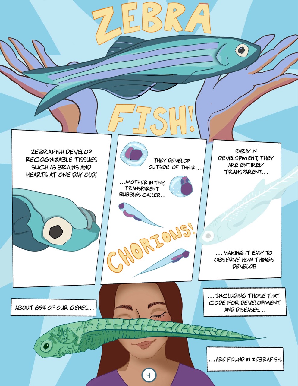
Panel 1: “Zebra Fish!” Two human hands with palms up raise up a larger-than-life zebra fish. “Zebrafish” is written in large block letters.
Panel 2: “Zebrafish develop recognizable tissues such as brains and hearts at one day old!” A small, blobby zebra fish with fully formed eyes.
Panel 3: “They develop outside of their mother in tiny, transparent bubbles called chorions!” A zebrafish develops inside the panel, going from an amorphous blob inside a bubble to a blob with recognizable eyes inside the bubble, to a small larval fish, to a fully adult zebra fish.
Panel 4: “Early in development, they are entirely transparent making it easy to observe how things develop.” A transparent zebrafish that allows you to see its internal structures including its spine.
Panel 5: “About 85% of our genes including those that code for development and diseases are found in zebrafish.” The dark-haired scientist faces forward with her eyes closed, a large zebrafish larvae obscures most of her face.
Page 5
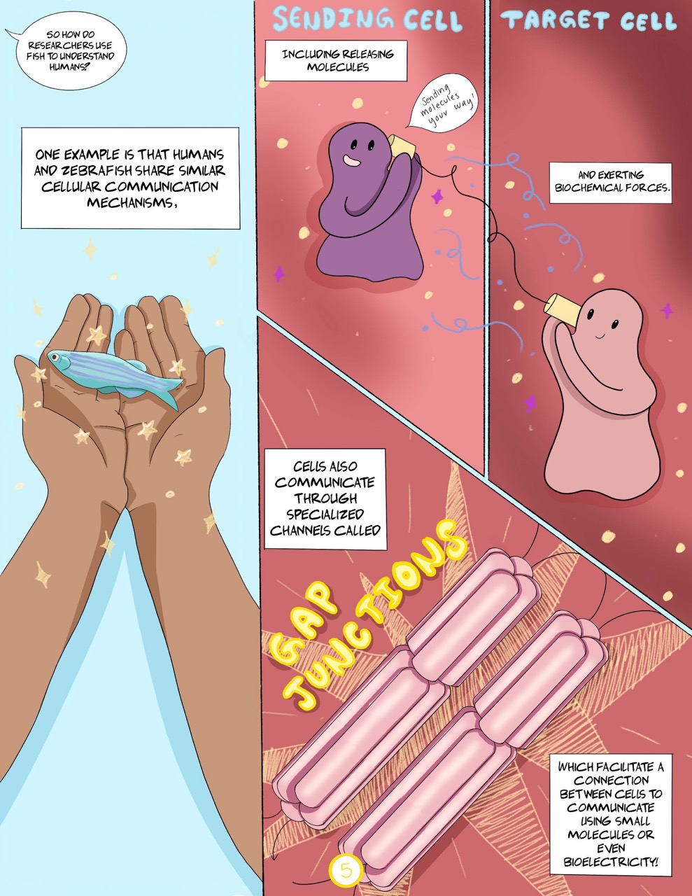
Panel 1: A long vertical panel. “So how do researchers use fish to understand humans?” The scientist cups a small zebrafish in her upturned hands. “One example is that humans and zebrafish share similar cellular communication mechanisms,”
Panel 2 and 3: “including releasing molecules and exerting biochemical forces.” In panel 2 an anthropomorphic cell, the “sending cell,” holds one end of a tin can phone and says, “Sending molecules your way!” In panel 3 the “target cell” listens to the other end of the tin can phone.
Panel 4: “Cells also communicate through specialized channels called gap junctions. Which facilitate a connection between cells to communicate using small molecules or even bioelectricity!” The words “gap junctions” are emphasized in block letters, and two gap junction structures are illustrated. They are segmented structures made up of long tubes connected in a circle.
Page 6
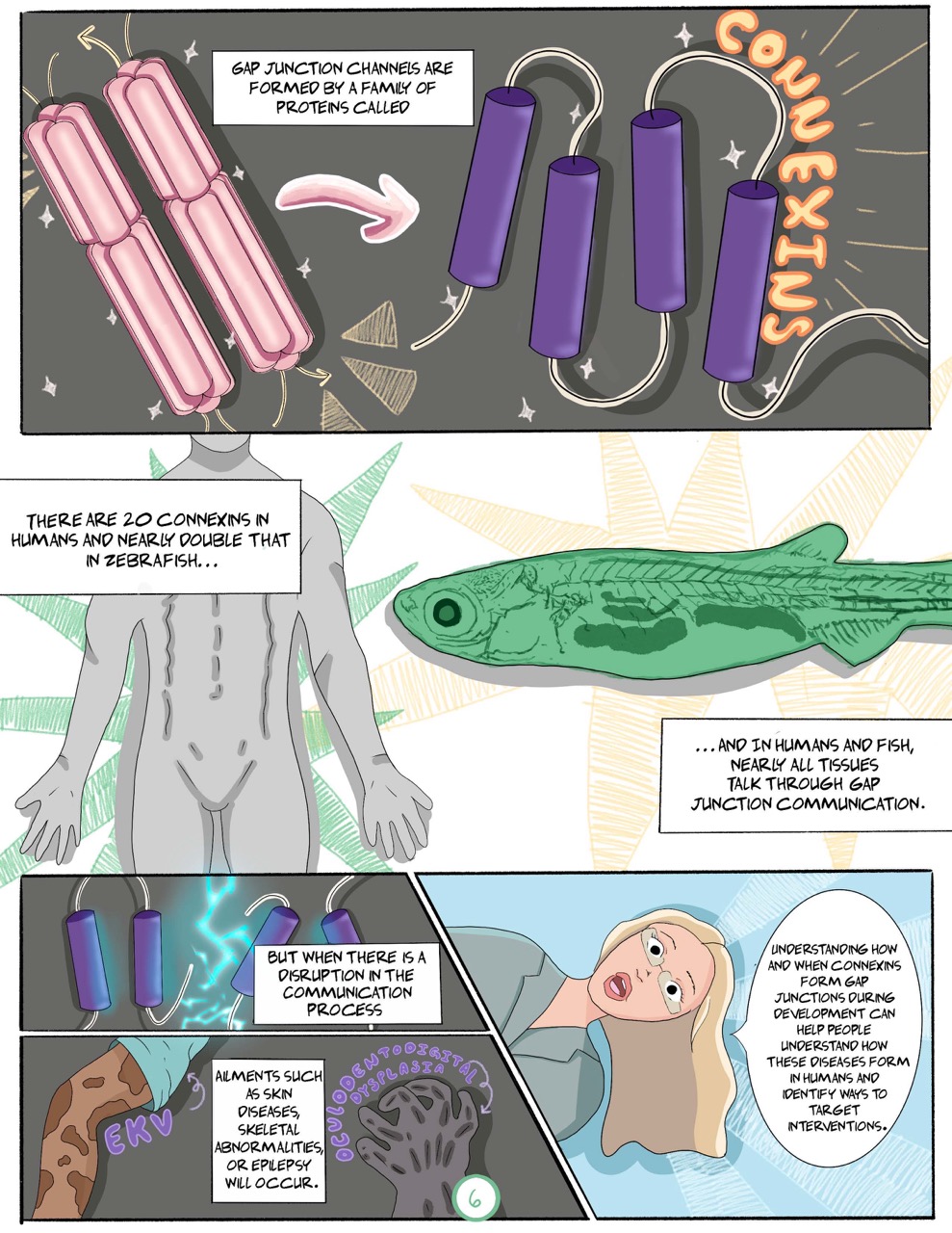
Panel 1: “Gap junction channels are formed by a family of proteins called connexins.” An arrow points between a diagram of gap junctions to a diagram of connexins. The cylindrical segments of gap junctions are made of connexins, which are cylindrical tubes connected by a thin string.
Panel 2: “There are 20 connexins in humans and nearly double that in zebrafish…” A diagram of a human body with defined muscles. Next to the person is a diagram of a zebrafish, its spine, internal structure, and organs are all visible through its skin. “…and in humans and fish, nearly all tissues talk through gap junction communication.”
Panel 3: “But when there is a disruption in the communication process” the string between a series of connexins is broken by a flash of lightning.
Panel 4: “Ailments such as skin diseases, skeletal abnormalities, or epilepsy will occur.” A human leg with discolored patches of skin is labeled “EKV.” A hand with grey skin and dark grey patches is labeled “Oculodentodigital dysplasia.”
Panel 5: The blonde scientist leans sideways into the frame, “Understanding how and when connexins from gap junctions during development can help people understand how these diseases form in humans and identify ways to target interventions.”
Page 7
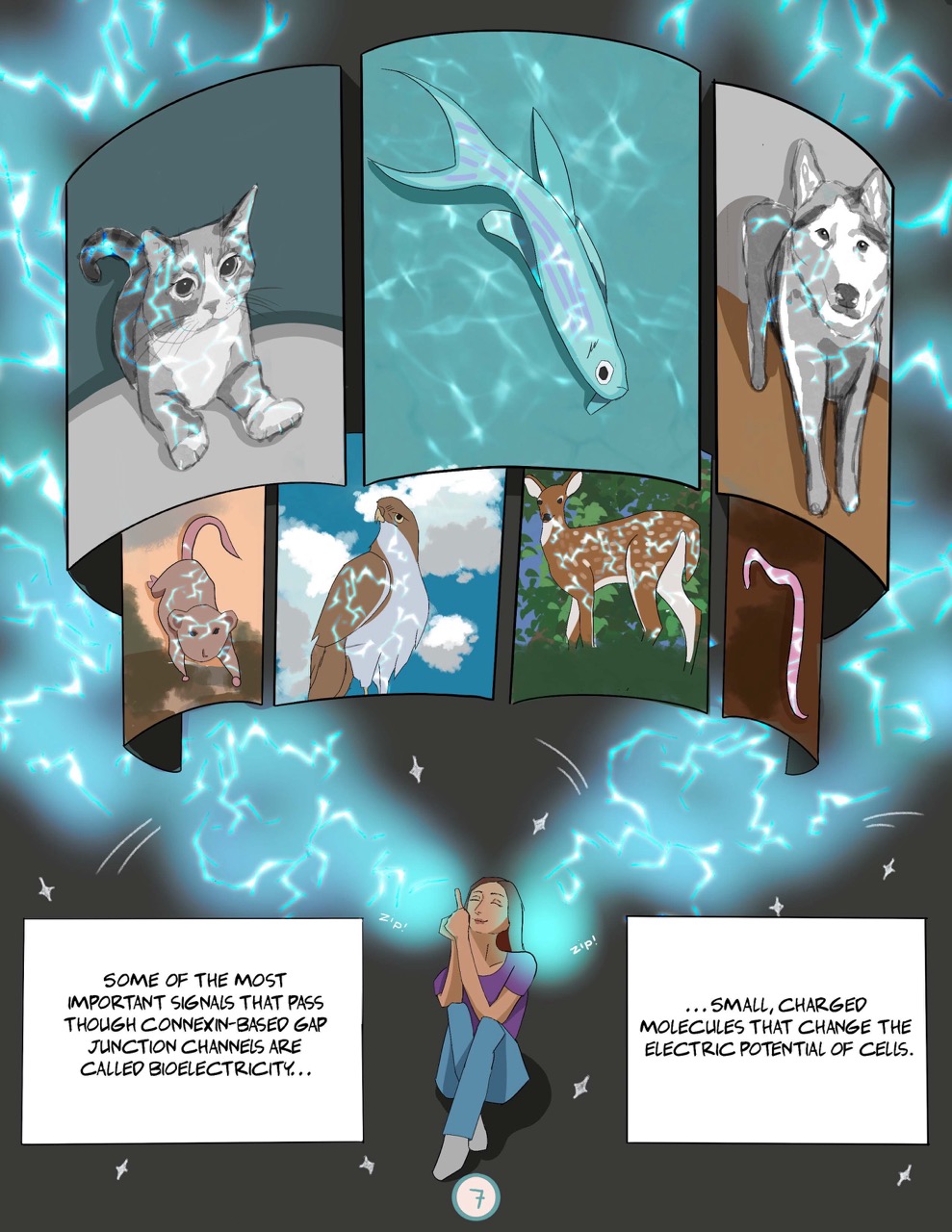
Full page spread: “Some of the most important signals that pass through connexin-based gap junction channels are called bioelectricity. Small, charged molecules that change the electric potential of cells.” Near the bottom of the page a young woman sits with crossed legs pointing upward. Above her head are a series of panels. Around the panels and overlayed withing the panels is bluish white bioelectricity lightning. Each panel has a different animal in it, from left to right the animals are: a small cat, a zebrafish, a husky, a mouse, a large hawk-like bird, a deer, and a nematode.
Page 8
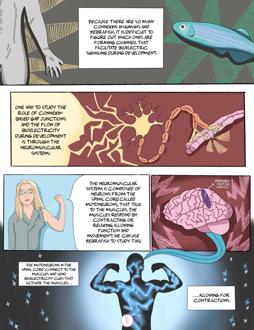
Panel 1: “Because there are so many connexins in humans and zebrafish, it is difficult to figure out which ones are forming channels that facilitate bioelectric signaling during development.” On one side of the panel is a person’s torso, on the other side is a zebrafish.
Panel 2: “One way to study the role of connexin-based gap junctions and the flow of bioelectricity during development is through the neuromuscular system!” A diagram of a neuron: a star shaped nucleus with branching dendrites and a long axon covered in a protective myelin sheath. At the end of the axon are branching tendrils with synapses that are connected to a muscle fiber. At one of the ends of a synapse the location of a gap junction is highlighted.
Panel 3: The blonde scientist has ripped one of the sleeves off her shirt, she flexes her bicep. “The neuromuscular system is composed of neurons from the spinal cord, called motoneurons, that talk to the muscles. The muscles respond by contracting or relaxing, allowing function and movement! We can use zebrafish to study this!”
Panel 4: “The motoneurons in the spinal cord connect to the muscles and send bioelectricity cues that activate the muscles, allowing for contractions.” In the center of the panel the silhouette of a person’s upper body flexes both arms. A network of nerves all lit up with bioelectricity run through the person’s body. An arrow points from their head to a sub-panel with a diagram of a brain. An oval in the upper-center of the brain is labeled “primary motor cortex.”
Page 9
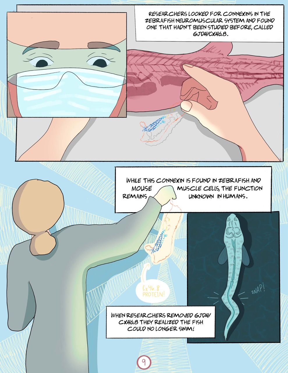
Panel 1: A close-up on the blonde scientist’s eyes and lower face. She wears a surgical mask and clear safety glasses.
Panel 2: This panel is larger and partially obscured by the first panel. It shows a POV shot of the scientist’s hands hovering above a larger-than-life zebrafish body. In one hand she holds onto a colorful structure made up of many thin wiggly lines. “Researchers looked for connexins in the zebrafish neuromuscular system and found one that hadn’t been studied before, called GJD4/CX46.8.”
Panel 3: The scientist holds up the wiggly structure, labeled “CX46.8 protein!” “While this connexin is found in zebrafish and mouse muscle cells, the function remains unknown in humans. When researchers removed GJD4/CX46.8 they realized the fish could no longer swim!” An inset panel shows an overhead view of a zebrafish. There is a sharp bend in its tail accentuated by emphasis lines and a “snap!”
Page 10
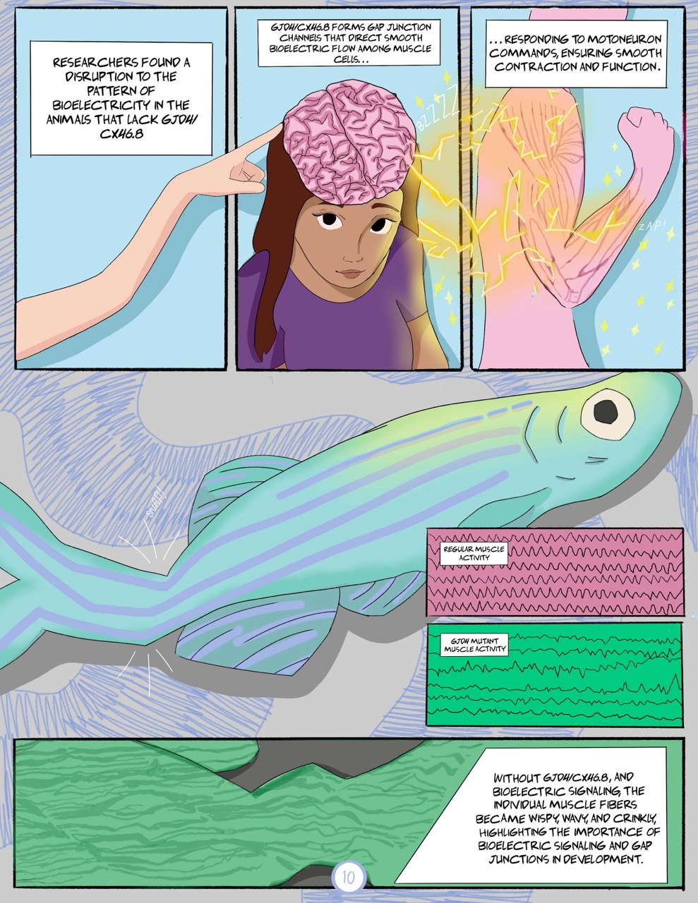
Panel 1 and 2: “Researchers found a disruption to the pattern of bioelectricity in the animals that lack GJD4/CX46.8” The blonde scientist reaches through the panel into panel two. In panel two the other scientist looks up at the reader. The top of her head has been removed so you can see her brain. The blonde scientist points to the side of her brain. “GJD4/CX46.8 forms gap junction channels that direct smooth bioelectric flow among muscle cells…”
Panel 3: “Responding to motoneuron commands, ensuring smooth contraction and function.” Bioelectric lightning buzzes from the brain in panel two into panel three. The bioelectricity arcs into a diagram of a person flexing the muscles in their arm. “Zap!”
Panel 4: A borderless panel that takes up the whole width of the page, an iridescent zebrafish fills the panel. It has a crick in its tail “snap!” Two subpanels show “regular muscle activity” and “GJD4 mutant muscle activity.” The regular muscle activity is a series of lines with even peaks and valleys. The GJD4 muscle activity shows irregular lines with shallow and sharp peaks and valleys.
Panel 5: “Without GJD4/CX46.8, and bioelectric signaling, the individual muscle fibers became wispy, wavy, and crinkly, highlighting the importance of bioelectric signaling and gap junctions in development.” A long narrow panel showing a closeup of zebrafish muscle fibers looking wavy.
Page 11
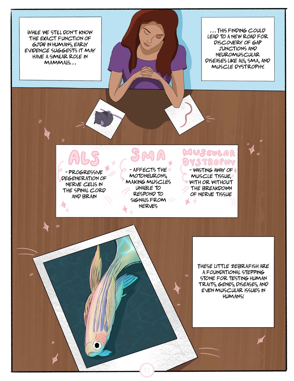
Full page spread: “While we still don’t know the exact function of GJD4 in humans, early evidence suggests it may have a similar role in mammals. This finding could lead to a new road for discovery of gap junctions and neuromuscular diseases like ALS, SMA, and muscle dystrophy.” Seen from above, the dark-haired scientist sits at a large wooden table, looking at pictures of a mouse and a nematode.
“ALS: progressive degeneration of nerve cells in the spinal cord and brain.”
“SMA: affects the motoneurons, making muscles unable to respond to signals from nerves.”
“Muscular dystrophy: wasting away of muscle tissue, with or without the breakdown of nerve tissue.”
A polaroid picture of a zebrafish sits on the table at the bottom of the page. “These little zebrafish are a foundational stepping stone for testing human traits, genes, diseases, and even muscular issues in humans!”
Page 12
“For more on Bentley Smallwood’s work.”
“Thank you for reading!”
“For more on Dr. Rachel Lukowicz-Bedford’s study.”
The page is decorated with stars, hearts, a husky dog, and a grey cat.
About the authors
name: Bentley Smallwood

name: Rachel Lukowicz-Bedford, Ph.D.
institution: University of Oregon
website: https://www.adammillerlab.com/